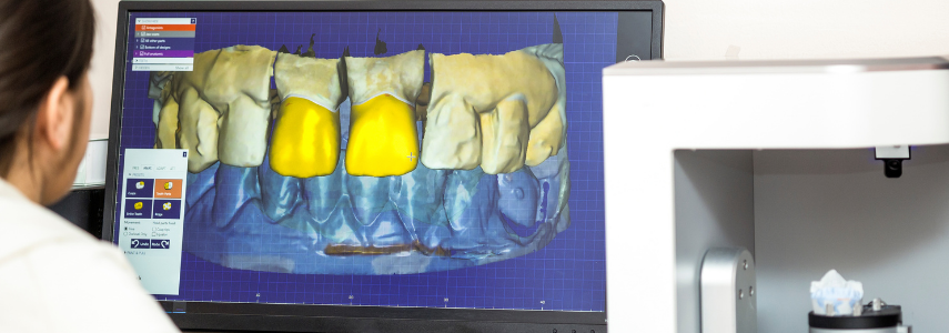
Technology has changed dentistry with great benefit to the patient; in addition to making surgery less invasive, it also improved the effectiveness of diagnosis as in the case of the use of the intraoral scanner for the detection of interproximal caries.
How does the intraoral scanner for the detection of interproximal caries work?
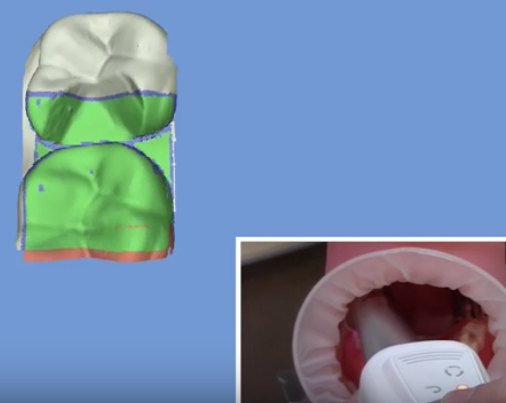
The intraoral scanner uses the NIR infrared light transillumination method which makes it easier to identify occlusal and interproximal carious lesions.
When the NIR transillumination is emitted on the buccal and lingual gingiva of the examined teeth, it is possible to observe the alveolar bone and dental tissues through a high-definition camera.
The images that the dentist can see through the camera allow an effective diagnosis thanks to the light reflection modes.
Intact enamel is visualized with a light gray color because NIR light is easily transmitted through this tissue, while in the presence of dentin and demineralized tissues the color appears dark gray.
By calibrating the wavelength of the NIR illumination, a contrast can be obtained that clearly distinguishes interproximal caries from healthy dental tissues.
Intraoral scanner and conventional methods: comparison of diagnostic results
In a recent research, published in the Journal of Dentistry, the use of intraoral scanners with infrared transillumination, NIR for the detection of interproximal caries lesions and conventional methods of caries detection were compared.
The sample examined was composed of 95 permanent posterior teeth divided as follows:
- Group 1 – diagnosis of interproximal caries using a prototype tip working with the intraoral scanner system with NIR light emission;
- Group 2 – diagnosis of interproximal caries using DIAGNOcam;
- Group 3 – diagnosis of interproximal caries by visual and radiographic examination
The results of the research
All methods showed excellent reliability, two independent examiners evaluated the NIR images obtained with both intraoral scanners.
As for the initial carious lesions, the diagnosis with an intraoral scanner was equal to or better than the radiographic evaluation and clearly superior to the visual examination. The radiographic diagnosis and evaluation with intraoral scanner was similar in detecting moderate-extensive dentinal carious lesions, while the visual examination showed a lower value.
It can be concluded that the use of intraoral scanners with NIR transillumination guarantees a good overall diagnostic performance for the detection of interproximal caries.


